


From childhood, we are used to running, dancing, boys want to climb and play football, girls in ropes and much more.And the active lifestyle is going so much into the human mind that over the years, when the muscle withdrew, somewhere united became ill, the person does not even pay attention to-"Well, you think how many times Kolenko hurt."Here in today's article we will talk, and why the knee can damage, and if this is always the usual result of sharp movement.
What is arthrosis?
arthrosis -A group of diseases of the skeletal muscle system of different origin, but with similar biological, morphological and clinical manifestations.The basis of their development is the degenerative lesion of all the ingredients of the knot, mainly cartilage, small bone, synovial membranes, ligaments, capsules and periarticular muscles, with the formation of marginal osteophytes and a clearly hidden synovitis.Since with this disease pathological changes capture both cartilage and bone tissue.
Arthrosis is often called osteoarthrosisAnd sometimes osteoarthritis.
Statistics (Epidemiology)
Among all joints of the joints, arthrosis is up to 80% of cases.
The disease develops mainly in middle and old age.At a young age, arthritis can occur after damage to the joints, inflammatory processes, and with congenital pathology of the muscular system.
Signs of X -Rray arthrosis are detected in most people over the age of 65 and almost 95%over 70 years old.
Women suffer from arthritis almost twice as often as men.The incidence rate increases during postmenopausal.
A major role in the development of arthrosis is played by hereditary factors.It is proven that the frequency of developing the disease in the families of patients with osteoarthritis is twice as high as in the population as a whole, and the development of arthrosis in people with congenital defects of the muscular-Skeletal system increases by 7-8 times.
Arthrosis - ICD
- MKB-10: M15-M19, M47
- MKB-9: 715
- MKB-9-Km: 715.3
Symptoms of arthrosis (clinical photographs)
The clinical manifestations of the disease and their severity depend on the localization of the pathological process, the state of the patient's health and the image of his life.
The first signs of arthrosis
Arthrosis often begins gradually, invisibly to the patient.
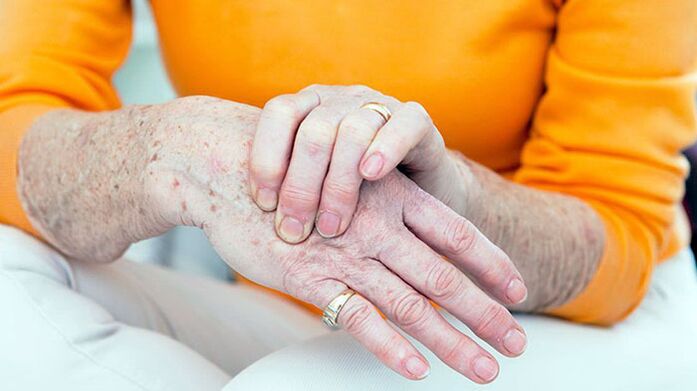
The first symptom of the disease is usually a short short -term joint pain (arthralgia), which holds the largest load.These are, first of all, the nodes of the lower extremities-the knee, hip, plus -phalanx of the first toe.From the joints of the upper limbs, the interphalngeal joints, the knot of the carpal finger pattern of the brush is most often affected.
Arthrosis usually begins with the lesion of a node, but after a while the other joints are involved in the process.
The main symptoms of arthrosis
With arthrosis, patients complain of pain, cramping, restriction of joint movements, swelling and joint deformation.
Separately, it is worth stopping in the nature of the pain.With arthrosis, mechanical and initial pain is possible.Mechanical pain appears with load on the affected joint.Such pain worries mostly in the evening at rest, disappears after a few hours of rest.The appearance of this type of pain is associated with a gradual increase in bone pressure during physical exercise.Pressure causes bone beams and irritation of painful bone tissue.
The onset of pain appears at the beginning of the walk, then quickly stops and occurs again during physical exercise.The onset of pain can occur with friction of the articular surfaces of the affected joint.Small necrotic cartilage particles fall on the surface of the cartilage.In the first steps, these particles are pushed into the joining bag cavity and the pain rests.
With arthrosis, the pain can be associated with periartrit and strain (inflammation of the periarticular soft tissue, ligamentous apparatus and articular bags).This pain occurs only during the movements in which the affected tendons, as well as in certain positions of the joint during the movements, participate.
Pathological changes, as a rule, begin with large joints, which are subject to large physical exercises during the day.At the beginning of the disease, the pain occurs as a result of the non -compliance with the possibilities of the microcirculation canal with the needs of the articular tissue.Therefore, to reduce pain, patients slowly take the first steps and only then accelerate the rhythm of walking.The pain can occur after half to two hours walking or working in a standing position.This is a signal to change load, short -term rest or type of work.
In the later stages of the disease, arthralgia can occur with minimal loads on the joint and remain at rest for a long time.This is due to the fact that in later stages, rude changes in joint tissue, articular cartilage destruction and secondary synovitis are formed.With the development of massive, gross changes in bone tissue, its individual fragments can be separated and, falling into the joint gap, cause a sharp pain.This phenomenon is called the symptom of the common mouse.
During examination of the joints, deformation is important.In addition, with arthrosis, there is a thickening of the periastal soft tissue, hypotrophy of the regional muscle, the displacement of the axis of the limbs.The obesity of bone -growing interphalangeal joints and the seal of periarticular fabrics is called Gerberden joints.
Pain when it feels that the knot is localized in the joint gap, places of joint capsule attachment, but this symptom of the disease is not always.Ellingance and joint pain is determined by secondary synovitis.
Violation of common function in the early stages of arthrosis manifests itself with a restriction of amplitude of movements.This is due to periosematic tissue lesion and synovitis.
In the later stages of the disease, the clinical manifestations of contractions take place different in terms of severity.Most often, knee and hip joint functions are damaged.
Symptoms of arthrosis depending on the localization of the pathology
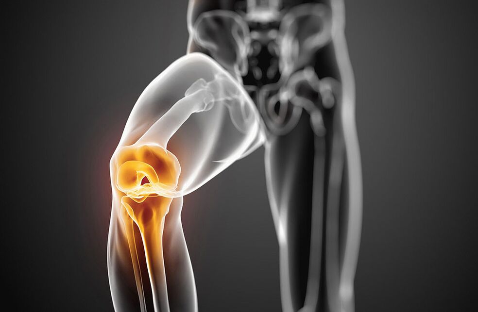
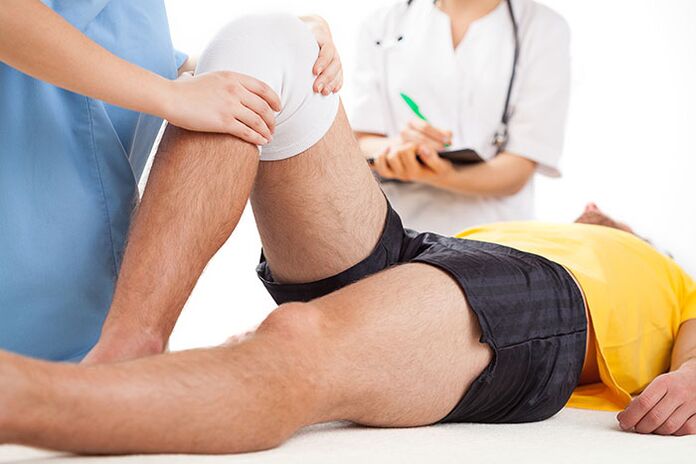
Arthrosis with damage to the knee joints - symptoms
The lesion of the knee joints with arthritis is called gonarthrosis.Primary gonartrosis develops in women in menopause.The reasons for the secondary are most commonly the knee joint injuries and a violation of static with spinal cord bending, flat feet.Patients complain of pain in the knee joint that occurs during movements, especially when walking up the stairs.The pain is localized to the front or inside the knee joint.The movements in the union are limited: the first bending, and the subsequent extension.When you move, a cramp often occurs.With the development of reactive synovitis, pain during movements intensifies and disturbed at rest.The swelling of the joint, pain during palpation, redness (hyperemia) and an increase in skin temperature are determined.Over time, due to bone growth, the deformation of the knee joints occurs.
Arthrosis with damage to hip joints - symptoms
The hip joint lesion is called coxartrosis.This is the most severe form of arthrosis.The causes of the disease can be congenital dysplasia of the hip joints, injuries, menopause.Patients have pain in the joints during movements, in a standing position.The limitation of movements in the joint is gradually increasing (the first internal and outer rotation, subsequent flexion).There is a madness associated with the shortening of the limbs.With bilateral damage, the walking of the ducks is typical.The atrophy of the thighs and buttocks develops.No swelling of the joints with cartridges.It perfectly defines limited pain in the femur's head.
In the initial stage of arthrosis, the common functions are preserved.With the further development of the disease, it is first temporarily limited, and then the ability to work is completely lost, the patient loses his self -care ability, needs external help.
Causes of arthrosis
Arthrosis is based on the primary degeneration of the articular cartilage with the devastating associated changes in bones that form the joint.Such degeneration occurs as a result of an imbalance between mechanical loads on the joint surface of the cartilage and the possibility of compensation for this load.
In developing degenerative changes in articular cartilage, several factors may participate at the same time:
- Functional overload, including professional, housewives and sports, causing cartilage mycotrauma;
- joint injuries;
- infectious and non -specific inflammation of the joint;
- joint dysplasia, leading to a violation of comparison of common surfaces;
- Violation of body static as a result of curvature of the spine (kyfosis, scoliosis, pathological lordosis, etc.), flat feet;
- Chronic hemartrosis:
- disease with metabolic disorders (gout, overweight, condrocalcinosis);
- Osteodystrophy or lecturer's disease;
- osteomyelitis;
- Pathology of the peripheral nervous system with loss of sensitivity;
- Endocrine pathology (acromegaly, diabetes, amenorrhea, hyperthyroidism);
- Hereditary tendency.
Arthrosis risk factors include old age, female sex, overweight.
Development mechanism
Disordishes metabolic disorders in the cartilage are based on quantitative and qualitative changes in the main cartilage substance.The main substance consists of proteoglycans that provide collagen stability.Arthrosis development is associated with insufficient formation or increased destruction of cartilage components.
With osteoarthritis in cartilage tissue, hyaluronic acid content, chondroitin and keratin decreases.In addition, changed proteoglycans lose the ability to keep water.It is absorbed by a collagen that swells, causing a decrease in cartilage resistance.
If chondrocytes are damaged, they begin to produce collagen and proteoglycan non -characteristic of normal cartilage tissue.These altered substances cause the loss of biochemical properties of the cartilage.
Of great importance in the development of arthrosis are immune disorders.The destruction of cartilage proteinoglycans is associated with the emergence of cellular and humoral immune reactions.On the other hand, this causes progressive fibrosis and sclerosis of the synovial membrane, pathological changes in the intraarticular synovial fluid and a dishersement of the cartilage.The inferior synovial shells supports the advancement of degenerative changes in the joint cartilage.
An inheritance factor has a certain value in the development of arthrosis.
Classification of arthrosis
Arthrosis is divided into two groups: primary and secondary.
In distribution (primary arthrosis):
- Local (with damage to three nodes)
- Common or generalized polycartrosis (loss of three nodes or more).
Subject to destination (secondary):
- A. tasobed union (corsartrosis);
- A. knee joint (gonartrosis);
- A. elbow node;
- A. shoulder joint;
- A. back;
- A. Cervical Department (Uncoarthrosis);
- A. hands;
- A. ankle connection (cruzartrosis)
- A. Stop.
From etiology:
- post -traumatic
- metabolic
- Due to endocrine pathology.
Diagnosis of arthrosis
The variety of clinical manifestations and arthrosis variants makes it difficult to diagnose the disease early.The falsity of the diagnosis is also associated with the lack of specific symptoms, the hidden onset of the disease.Of great importance is the definition of factors that contribute to the development of arthrosis:
- chronic joint trauma;
- long -term execution of stereotype movements;
- physical activity in union for a specified time;
- violation of salt or fat metabolism;
- Hereditary vice of the muscular -Skeletal system.
An X -Ray examination is of the most important meaning in the diagnosis of arthrosis.A viewing radiograph of both knee joints is performed in a direct position, a bent position, in addition in a side position.Classic signs of arthrosis in radiographs are: narrowing of the joint gap, the presence of osteophytes, subcondrial bone sclerosis and subcondular cysts.There are the following stages of radiological changes in arthritis:
- 0 - No changes.
- I - Suspicious radiological signs.
- of secondment - Minimal changes (slight narrowing of the joint gap, auxiliary osteosclerosis, single osteophytes).
- III - moderate manifestations (moderate narrowing of the statute, numerous osteophytes).
- Iv- Expressed changes (the common gap is not visible, multiple rude osteophytes are determined), synovitis is often present.
In the presence of these symptoms, further tools are not necessary.
In low absence or severity, joints, MRI, skintigraphy are performed.
Clinical blood tests, urine and intraarticular synovial fluid are not included in the list of mandatory studies for arthrosis diagnosis.But these tests are needed to exclude such articular pathologies.
Main clinical and diagnostic signs of arthrosis:
- Mechanical joint pain;
- fatigue;
- a feeling of instability in the nodes of the lower extremities;
- damage to the joints of the first toe and hands;
- gradual onset of the disease;
- slow progressive current;
- joint deformation;
- Regional muscle hypotrophy;
- repeated synovitis;
- restriction of movements in union;
- X -Ray changes.
Arthrosis should be differentiated with damage to joints with rheumatoid arthritis, infectious, metabolic and reactive arthritis.
Rheumatoid arthritis, unlike arthritis, begins with inflammation of the small joints of the hands and feet.It is characterized by intense inflammatory pain, breakfast stiffness of the joints, the presence of rheumatoid joints.
Gotric arthritis is found mainly in men.High local activity with acute paroxysmal pain in the first plus-phalanx node of the big toe is characteristic.With the ground floor, the presence of the tofus is typical, there are "punch" radiographs.
Psoriatic arthritis is characterized by skin lesions, especially scalp, finger -shaped deformity and a bright skin raspberry over the affected joints.
Infectious arthritis is characterized by an acute onset, rapid development and course, sharp pain, high fever and effectiveness of antibacterial therapy.
The treatment of arthrosis
The treatment for arthritis should be long, complex.Basic principles of arthrosis treatment:
- Discharge of joints (accurate mode of mobility and mechanical loads, dose walking, reduction of body weight, exclusion of prolonged stay, wearing weights, strengthening muscle-ligamentous apparatus using physiotherapy exercises, massage, electrical stimulation).
- Conservative correction of static disorders (use of orthopedic, corset, supervisory shoes).
- Impact on general metabolism and blood circulation (use of biostimulants, vasodilating drugs, balneotherapy courses and physiotherapy twice a year).
- Elimination of reactive synovitis, anti -inflammatory therapy.
Arthrosis patients indicate a diet with a restriction of salt, sugar, strong tea, coffee, smoked meat, sharp vessels.This improves the sensitivity of vascular and articular receptors, restores the tone of blood vessels, normalizes the exchange in the chondrocytes.With arthritis, it is necessary to drink enough juice (at least 8 glasses of water a day).
The treatment of arthrosis drugs involves the use of anti -inflammatory and fast sedatives (non -inflammatory non -steroidal -nsaids), basic medicines -kukondroprotectors.Non-selective and selective TSO-2 inhibitors are used by NSAID.
As local therapy for affected joints, NSAIDs are used in the form of an oil or gel.
In the presence of reactive synovitis, tendinitis or tendovaginitis, when NSAIDs treatment is ineffective, proper intraarticular or intramuscular corticosteroid administration.
Basic chondroprotectors (chondroitin, glucosamine, hyaluronic acid) therapy is used to prevent the degeneration of the joint cartilage.
The treatment of chondroprotectors is indicated in the clinical and radiological stages of arthrosis I-III.
In addition to direct chondroprotectors, medicines that stimulate the restoration of cartilage tissue (biogenic stimulants) are used.These medicines are used during prayer, in the absence of reactive synovitis.
With arthrosis, medicines that improve microcirculation are also shown.In the presence of varicose veins of the lower extremities, the correction of the venous blood flow is needed.
In patients with arthrosis, it is necessary to diagnose and treat osteoporosis in a timely manner.
Physiotherapy of arthrosis
Physical methods of treatment are also associated with basic arthrosis therapy.Under their influence, metabolic processes are stimulated, microcirulation of blood and tissue fluid, neurogumor regulation is restored.
The arthritis treatment complex includes inductor, microwave therapy, pulsating currents, medication electrophoresis and magnetotherapy.To eliminate the synovitis, the ultraviolet radiation of the area of the joints affected in erythema doses, an ultra -high electric field, electrophoresis with analgin, dual or hydrocortisone is used.
To prevent arthrosis progression, it is recommended to reduce body weight, avoid increasing loads on the joints, walking in a suppressed area, increases moisture and hypothermia.An individual selection of shoes and supervisors is important.
With gonartrosis, regular physical exercises, swimming, cycling is shown to strengthen the muscles.Severe and light athletics classes, football is not recommended.
Therapeutic exercises are performed differently, in the sitting position, lie, in the pool.The movements should not be intense, traumatic, their volume and the number of repetitions are gradually increased, avoiding overloads.
Popular and effective methods of treatment of arthrosis also include massage and kine or or.
With significant changes in joints with deformation, mobility restriction, surgical treatment is recommended.Arthroplastics, endoprosthetics, osteotomy are performed.
The prognosis of the disease
Primary arthrosis rarely leads to complete disability.In the presence of reactive synovitis, patients are temporarily become disabled, and sometimes they are forced to change their profession.With secondary coconut, the prognosis is less favorable due to the rapid progressive course of the disease with the development of an important damaged joint function.In such cases, disability can occur during several years of illness.
Prevention of arthrosis
Primary prevention of arthritis should begin in childhood.Is as follows:
- prevention and treatment of scoliosis;
- Correction of flat feet using special supervisors;
- the class of physical education to strengthen muscles and ligaments;
- rational nutrition and prevention of metabolic disorders;
- restriction of heavy sports in childhood and adolescence;
- alternative work sitting on a table with walking;
- Accurate work organization and the rest of the employees in enterprises where there is heavy physical activity.
Secondary prevention provides for measures that prevent the development of repeated reactive synovitis.These include dose walking, restricting physical exercise, walking with support and other measures that discharge the joints.With severe arthrosis symptoms, it is necessary to consistently take underlying medicines.General strengthening therapy, blood circulation improvement and metabolism are recommended, annual bath treatment is recommended.
Which doctor will go to?
- Rheumatologist
- Orthopedic